Nuclear Mechanics and Mechanotransduction (Co-PIs: Luxton and Starr)
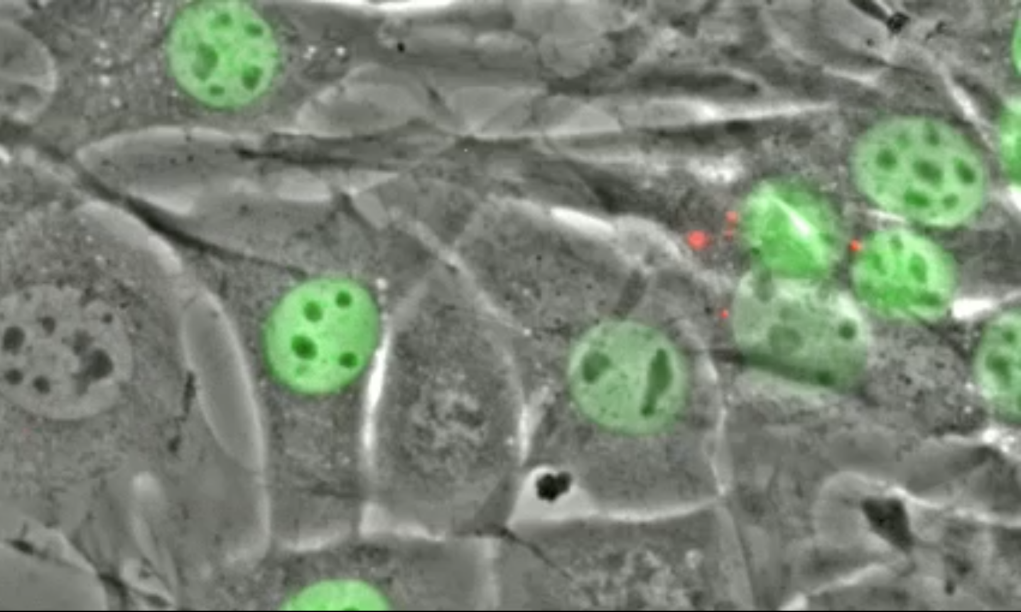
Defective transduction of mechanical signals into cellular responses is associated with human diseases, including cancer, cardiomyopathy, muscular dystrophy, neurodegenerative disorders, and premature aging. To understand how cells regulate their mechanical properties, we need to understand the molecular mechanisms of LINC (linker of nucleoskeleton and cytoskeleton) complexes, which span the nuclear envelope and mechanically integrate nuclei with the rest of the cell and its external environment. Our studies combine hypothesis-driven experiments with whole-genome CRISPR screens to identify new players in LINC complex-dependent mechanotransduction. Our goals are to uncover novel mechanisms for how LINC complexes regulate the cell. We study how LINC complexes are assembled. We aim to develop a small-molecule inhibitor of LINC complexes that will enable studies of LINC complexes in a wide variety of physiological and disease models. We will also identify mechanisms and new players in how nucleoli are regulated. Finally, we are exploring how LINC complexes regulate the mechanical properties of cells. We are developing new tools to study cellular mechanics in living tissues. LINC complexes play key functions in addition to mechanotransduction, including nuclear positioning, homolog pairing in meiosis, DNA damage repair, wound healing, spermatogenesis, and nuclear pore complex assembly. Our research is relevant to public health because mutations in LINC complex proteins are implicated in a wide range of diseases including ataxia, cardiomyopathy, hearing loss, muscular dystrophy, neurodegenerative disorders, Progeria, sterility, and various cancers. Thus, our studies are significant for understanding the molecular underpinnings of a wide variety of cellular and developmental processes as well as human disease pathogenesis.
Mechanisms of Nuclear Migration (PI: Starr, funded by NIH MIRA Grant)
Nuclear migration and anchorage are central to many cellular events. The Starr lab has uncovered a conserved network of nuclear envelope proteins and force generators that mediate nuclear positioning. LINC (linker of nucleoskeleton and cytoskeleton) complexes maintain nuclear envelope architecture, mark the surface of nuclei distinctly from the contiguous ER, and were instrumental in the early evolution of eukaryotes. We address the following using primarily C. elegans as a model: (1) How is the developmental switch between nuclear migration and anchorage mediated? We hypothesize that different LINC complexes are required for a nucleus to switch from migrating to being anchored. We further hypothesize that LINC directly interacts with the outer nuclear membrane to optimize the transfer of forces across the nuclear envelope. (2) How are nuclei anchored in large syncytial cells? It is important for nuclei to be evenly spaced so that multi-nucleated syncytia are able to act as a single unit. ANC-1 anchors syncytial nuclei, ER, and mitochondria through unknown, LINC-independent mechanisms, and hypothesize that ANC-1 organizes the cytoplasm through crosslinking the cytoskeleton. (3) How do nuclei favor one microtubule motor over another at different stages of development? The KASH protein UNC-83 mediates nuclear movements toward plus or minus ends of microtubules at different stages of development. We hypothesize that the choice is regulated by alternative isoforms of UNC-83 that differentially activate kinesin-1 motor activity. (4) How do nuclei deform to migrate through narrow spaces? Our data support a model where LINC complexes function parallel to branched actin networks to deform nuclei as they squeeze through narrow constrictions. We can view live nuclei throughout C. elegans development, including a tissue where nuclei migrate through narrow constrictions as a normal part of development. To complement our C. elegans genetic approaches, we collaborate to confirm our findings in mammalian tissue culture cells and an in vitro microtubule motor assay with TIRF microscopy. Our studies are expected to determine how LINC complexes are regulated at molecular level, how the outer nuclear membrane is involved in force transmission, how giant KASH proteins organize the global cytoskeleton and position organelles, how UNC-83 mediates the choice between dynein and kinesin-directed nuclear movements throughout development, and how actin helps nuclei squeeze through constricted spaces.
TorsinA-mediated regulation of cdc42 signaling and DYT1 Dystonia (PI: Luxton, funded by NIH R01)
DYT1 dystonia is a devastating neurological movement disorder characterized by uncontrolled muscle contractions that result in abnormal, involuntary postures. DYT1 dystonia is caused by a deletion (Δgag; ΔE) in the Tor1A gene encoding the luminal ATPases associated with various cellular activities (AAA+) protein torsinA. How the ΔE mutation causes DYT1 dystonia remains unclear because the basic cellular function performed by torsinA is unknown. This proposal seeks to close these critical gaps in knowledge, which will enable the rational design of urgently needed targeted therapies. Our recent work suggests that torsinA is i) required for the transduction of mechanical signals from the cytoskeleton into the nucleoplasm via the nuclear envelope spanning linker of nucleoskeleton and cytoskeleton (LINC) complex; ii) the activation of signaling mediated by the Rho GTPase Cdc42; iii) as well as proper axon outgrowth and growth cone morphology. Importantly, axonal tract disruptions that correlate with clinical severity are observed in the brains of DYT1 dystonia patients and the both the LINC complex and Cdc42 mediate axon elongation and guidance. Thus, we hypothesize that defective torsinA-dependent mechano-chemical signal transduction contributes to DYT1 dystonia pathogenesis. We will test this hypothesis in cultured mammalian cells and the African clawed frog Xenopus laevis using an array of established and novel biochemical, biophysical, fluorescent Rho GTPase biosensors, quantitative imaging and proteomics, as well as synthetic biological approaches. In this proposal, we will define how torsinA and its co-activator, the inner nuclear membrane protein LAP1, regulate the assembly of functional LINC complexes in cultured mammalian cells (Aim 1). We will determine how torsinA and its other co-activator, the outer nuclear membrane protein LULL1, control the activation of Cdc42 in cultured mammalian cells (Aim 2). Finally, we will test the role of torsinA-dependent mechano-chemical signal transduction during axon outgrowth in cultured X. laevis neurons and in the brains of living embryos (Aim 3). The results of these Aims will provide invaluable mechanistic insights into the emerging role of the nuclear envelope as a signaling node in development and disease. Furthermore, they will lay the foundation for the future development of novel therapeutic strategies for the treatment of other forms of dystonia, dystonia plus syndromes in which dystonia can occur in conjunction with another neurological disorder such as Huntington’s and Parkinson’s diseases, as well as other neurologic and neuropsychiatric diseases caused by mutations in LINC complex proteins including autism, ataxia, bipolar disorder, dementia, and schizophrenia.
Gordon and Betty Moore Foundation Scialog Award: Reconstructing time-resolved single-cell genome organization (Co-PI: Luxton)
The long-term goal of our GBMF project, which is a collaboration between myself and Drs. Brian Liau (Harvard University) and Bin Zhang (MIT), is to understand how the genome is organized in space and time within the nucleus as well as how this organization is disrupted in HGPS. Despite its importance, few general methods exist to measure 3D genome structure over time. Thus, we will develop strategies for fluorescently labeling regions of chromatin at defined positions within the nucleus in order to subsequently image their dynamics in living control and progeroid cells. To derive the underlying DNA sequence of the labeled regions and put the dynamics into context, we will reconstruct a genome structure that best fits the imaging data. We will specifically label genomic regions that are located proximal to the nuclear lamina (i.e. lamina-associated domains (LADs)), since the nuclear lamina is highly perturbed in senescent and HGPS cells. Our project has three Aims: 1) To fluorescently label LADs in living cells; 2) Image in vivo LAD positioning and dynamics in control and HGPS cells; and 3) Integrate imaging data with modeling to reconstruct time-resolved single cell structures. We envision that this technology and approach can be broadly applied to study other nuclear ‘landmarks’ and domains.
