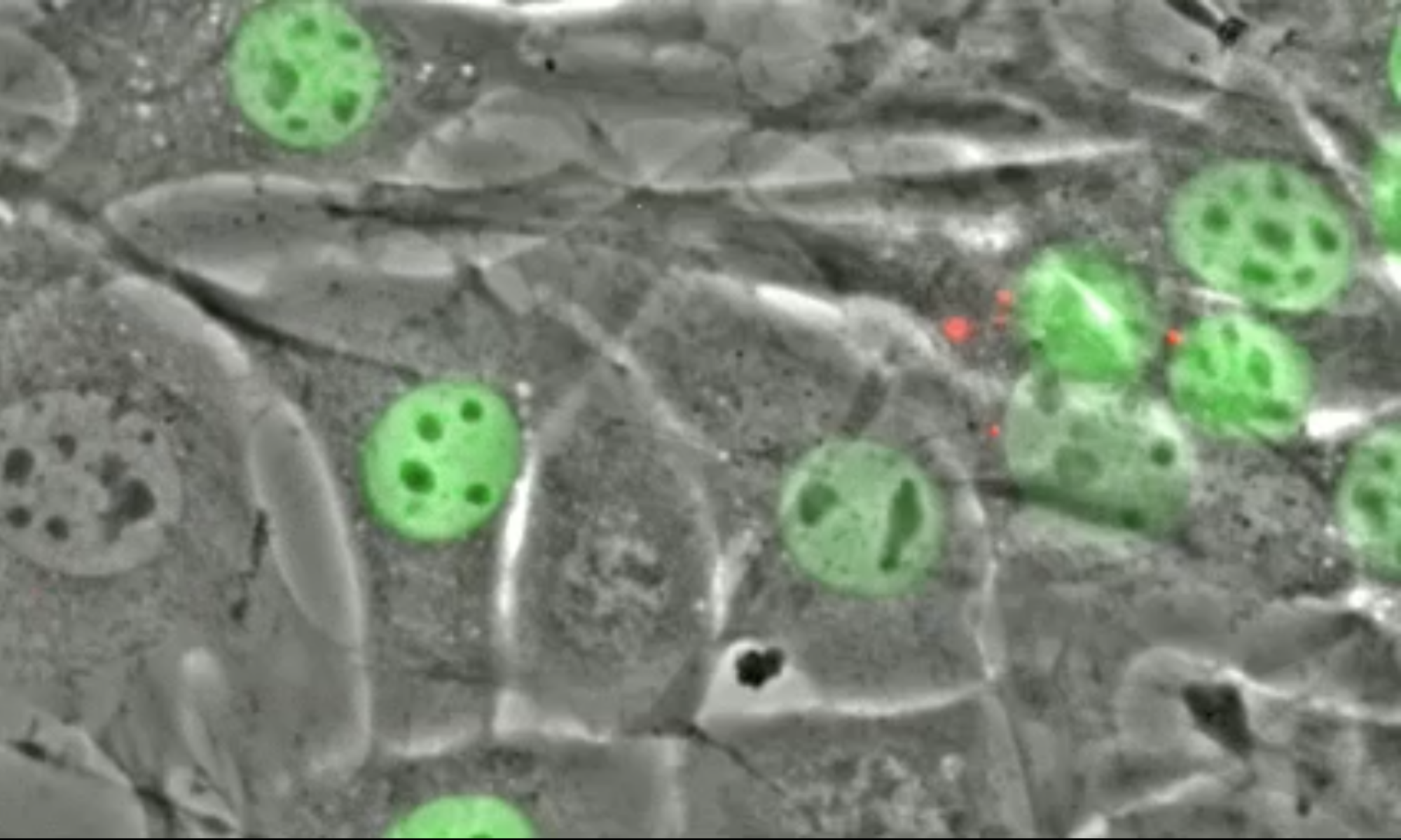
A ventral view of an L1 larvae expressing a tight junction::GFP (circles around P cells) and a GFP for fibrous organelles that connect muscles to the cuticle. For more details, see Bone et al 2016 Development.
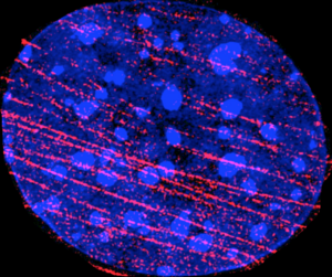
Super-resolution image (SIM, DeltaVision OMX) of a serum-starved wound-edge mouse embryonic fibroblast that was fixed in paraformaldehyde 60 minutes after being stimulated with LPA. Blue: DNA (DAPI). Red: Actin (rhodamine-phalloidin).
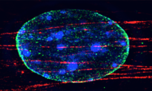
Super-resolution image (SIM, DeltaVision OMX) of a serum-starved wound-edge mouse embryonic fibroblast that was fixed in paraformaldehyde 60 minutes after being stimulated with LPA. Blue: DNA (DAPI). Green LINC complexes (anti- nesprin-2G). Red: Actin (rhodamine-phalloidin).
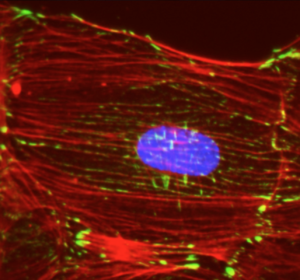
Epifluorescence image (Nikon) of a serum-starved wound-edge mouse embryonic fibroblast that was fixed in paraformaldehyde 60 minutes after being stimulated with LPA. Blue: DNA (DAPI). Green: focal adhesions (anti-vinculin). Red: Actin (rhodamine-phalloidin).
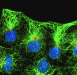
Epifluorescence image (Nikon) of serum-starved wound-edge NIH3T3 fibroblasts that were fixed in methanol 120 minutes after being stimulated with LPA. Blue: DNA (DAPI). Green: microtubules (anti-tyrosinated microtubules). Red: centrosomes (anti-pericentrin).
Live epifluorescence (left) and phase contrast (right) imaging (Nikon) of a serum-starved wound-edge Lifeact-mCherry-expressing NIH3T3 fibroblast stimulated with LPA. Images were taken every 5 minutes after LPA-stimulation.
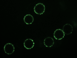
Spinning disk confocal (Olympus) image of full-length EGFP-tagged mouse SUN2 reconstituted in artificial nuclear membranes.
Pseudocolored epifluorescence image (Nikon) of abnormal nuclear envelope-associated lysine 48 (K48) poly-ubiquitination in tor1A/1B/2A/3A-knockout HeLa cells labeled by immunofluorescence.

A GFP marker is expressed in hypodermic syncytial nuclei. In this anc-1 mutant adult, the syncytial nuclei can be seen clustering. See Cain et al 2018 Current Biology for more details.
